


CARE & TREATMENT
 What are the symptoms of Glaucoma?
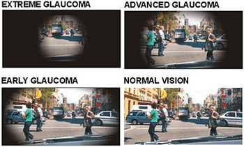
Glaucoma is known to be a silent thief of sight and hence has no symptoms. As Glaucoma affects the visual field rather than the sharpness of vision, patients may not notice any change in the vision in the initial stages. Inability to see objects on the side while walking or driving may be early symptoms of Glaucoma. Open angle glaucoma’s are mostly symptom free. Thus it is usually an incidental finding. Closed angle glaucoma patients may have occasional episodes of headache, pain, redness of eyes, blurring of vision, nausea and vomiting, and appearance of colored rings around lights during such episodes.
How can I confirm whether I have Glaucoma?
To confirm whether you have glaucoma you need to consult your Ophthalmologist (Eye doctor) who can tell you whether you have glaucoma. Diagnosis of Glaucoma requires very simple and basic eye examinations and tests and can be easily done in any reputed eye institute.
Can Glaucoma be cured?
Glaucoma, like diabetes or blood pressure cannot be cured permanently, but can be treated and kept under control. The vision lost due to glaucoma cannot be regained but can be retained. The aim of treatment is to delay the progression of the disease and preserve the present vision to out live the patient. Hence, it is important to diagnose the glaucoma in its initial stages and start treatment.
What are the treatment options for Glaucoma?
The basic aim of Glaucoma treatment is to control the pressure inside the eye. This can be done medically (either by reducing the production of the fluid inside the eye or by facilitating drainage of the same out o the eye), by LASERs and surgically (both of which facilitate the outflow of the fluid).
Is Glaucoma surgery or LASER procedure painful?
No, the surgery and the LASER procedure is painless and patients at the most feel a slight discomfort after the surgery
Is Glaucoma surgery a onetime affair like cataract surgery?
No, the success of Glaucoma surgery depends as much upon the follow up and post operative care as upon the skill of the surgeon. The follow up to the doctor’s clinic are more frequent after a Glaucoma surgery as compared to a cataract surgery, and may require small procedures during the follow up visit to maximize the outcome of the surgery.
Is Glaucoma preventable?
Although there is no scientifically proven method or medication to prevent Glaucoma, leading a healthy lifestyle and getting periodic eye checkups is a sure way to keep Glaucoma at bay.
How can you prevent blindness from glaucoma?
All those at risk must get their eyes examined regularly. All those diagnosed with the disease must follow-up with the doctors regularly and take timely treatment. Regular follow-ups and compliance with treatment is the way to prevent the blindness.
Lasers In Glaucoma
What is the laser treatment available for glaucoma?
Depending on the type of glaucoma you have various Laser procedures are available.
For angle closure glaucomas , a YAG Peripheral iridotomy is done, to make a small hole in the iris & open the drainage of the aqueous ( fluid in the eye) .
- For an acute angle closure attack, if a Yag PI does not work an argon laser is used to create small burns in the periphery to retract the iris from the angle, thus opening out the angle. This procedure is called laser trabeculoplasty.
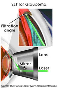
- In patients who have open angle glaucomas, primarily laser trabeculoplasty to open the clogged angles is done using either the argon laser (ALT) , better still the use of a Yag laser called Selective Laser trabeculoplasty (SLT) where minimal damage is done to the trabecular meshwork, & can be easily repeated. This will ensure flow of the aqueous and lower the IOP. It can be used as a primary mode of treatment.
- Post –operatively, if a deep sclerectomy has been done, the angle can be opened at a later date by goniopunctures with a Yag laser.
- If a Trabeculectomy has been done & the inner ostium is blocked by blood, iris etc, that too can be lasered post-operatively.
Selective Laser Trabeculoplasty or SLT, is a form of laser surgery that is used to lower intraocular pressure in glaucoma. It is used when eye drop medications are not lowering the eye pressure enough or are causing significant side effects. It may sometimes be used as initial treatment in glaucoma.
 SLT has been in use for 12 years in the United States and around the world.
This article is designed as a discussion to answer the most common patient questions and concerns regarding the procedure.
- Who is a candidate for SLT?
Patients who have open-angle glaucoma (the drainage system in the front part of the eye is open) and are in need of lowering of their intraocular pressure (IOP) are eligible for the procedure. Your eye doctor will make the final determination if you are a candidate. - How does it work?
Laser energy is applied to the drainage tissue in the eye. This starts a chemical and biological change in the tissue that results in better drainage of fluid through the drain and out of the eye. This eventually results in lowering of IOP. It may take 1month for the entire effect to appear. - Why is it called Selective?
The type of laser used has minimal heat energy absorption because it is only taken up by selected pigmented tissue in the eye. Sometimes it is referred to as a “cold laser.” Because of this, the procedure produces less scar tissue and has minimal pain. - What are the risks?
One key aspect of SLT is a favorable side effect profile, even when compared with glaucoma medications. Post-operative inflammation is common but generally mild, and treated with observation or eye drops or an oral non-steroidal anti-inflammatory drug - How effective is it?
SLT lowers the IOP by about 30% when used as initial therapy. This is comparable to the IOP lowering of the most powerful and commonly used class of glaucoma medication (prostaglandin analogs). This effect may be reduced if the patient is already on glaucoma medications. - How long does the effect last?
The effect will generally last between 1-5 years, and in rare cases, longer than that. If it does not last at least 6-12 months, it is usually not considered successful. - What happens if it wears off?
If SLT is effective at lowering IOP but this wears off over several years, the procedure can be repeated But the second treatment may not be as effective as the first and may not last as long. If SLT is not initially successful, repeat treatment is not likely to be effective. Alternatively, glaucoma medication can be used if the effect wears off over time. - What happens if it doesn’t work?
If SLT fails to lower the IOP, then the glaucoma is treated by other means such as medications or conventional surgery. The laser does not affect the success of these other types of treatment.
Will I still need to use glaucoma medications?
Some patients can be controlled with just laser treatment. Others require additional IOP lowering and may therefore need to use glaucoma medication as well. Think of the SLT as equivalent to one glaucoma medication. Just as some patients will require more than one glaucoma medication to control their IOP, some may also require laser plus one or more glaucoma medications. It is important to remember that SLT is not a cure for glaucoma, just as medication and surgery are not. Whatever method is used to treat glaucoma, appropriate follow up and testing with your eye care professional is critical.
Yag PI
Laser peripheral iridotomy is a treatment for narrow angle glaucoma. The surgeon uses an Nd:YAG laser to create a small hole in the peripheral iris. This improves the circulation of fluid inside the eye and widens the anterior chamber angle. Fluid which is produced behind the iris has easier access to the eye's internal drainage system. Sometimes this lowers the intraocular pressure, but that is not the primary goal of laser peripheral iridotomy. The primary goal of the procedure is to lessens the risk of acute angle-closure glaucoma.What to expect on procedure day
Once a YAG Pi has been scheduled, a nurse/ an assistant will administer some eye drops which will constrict your pupils. After 15 to 30 minutes, you will be escorted to the laser room, where the procedure will be performed. Your surgeon will use the Nd:YAG laser to place a small hole in the peripheral iris of the involved eye(s). During the procedure, you will see a brief flash of light, hear a clicking sound, and possibly feel a slight stinging sensation. The procedure will last only a few minutes. Your eye pressures will be checked shortly after the treatment, and you will be discharged home to resume your normal activities. Your doctor might prescribe some anti inflammatory eye drops to be used for a few days after the procedure. Patients occasionally experience transient mild redness, discomfort, light sensitivity, and blurred vision.Diagnostic tests for glaucoma
Early detection, through regular and complete eye exams, is the key to protecting your vision from damage caused by glaucoma. A complete eye exam includes five common tests to detect glaucoma.
It is important to have your eyes examined regularly. Your eyes should be tested:
- before age 40, every two to four years
- from age 40 to age 54, every one to three years
- from age 55 to 64, every one to two years
- after age 65, every six to 12 months
Anyone with high risk factors should be tested every year or two after age 35.
A Comprehensive Glaucoma Exam
To be safe and accurate, five factors should be checked before making a glaucoma diagnosis:
| Examining... | Name of Test |
| The inner eye pressure | Tonometry |
| The state and color of the optic nerve | Ophthalmoscopy (dilated eye exam) |
| The complete field of vision | Perimetry (visual field test) |
| The angle in the eye where the iris meets the cornea | Gonioscopy |
| Thickness of the cornea | Pachymetry |
Regular glaucoma check-ups include two routine eye tests: tonometry and ophthalmoscopy.
Tonometry
Tonometry measures the pressure within your eye. During tonometry, eye drops are used to numb the eye. Then a doctor or technician uses a device called a tonometer to measure the inner pressure of the eye. A small amount of pressure is applied to the eye by a tiny device or by a warm puff of air.
The range for normal pressure is 12-22 mm Hg (“mm Hg” refers to millimeters of mercury, a scale used to record eye pressure). Most glaucoma cases are diagnosed with pressure exceeding 20mm Hg. However, some people can have glaucoma at pressures between 12 -22mm Hg. Eye
pressure is unique to each person. Ophthalmoscopy
This diagnostic procedure helps the doctor examine your optic nerve for glaucoma damage. Eye drops are used to dilate the pupil so that the doctor can see through your eye to examine the state and color of the optic nerve.This is usually done in the same sitting on the slit-lamp with the help of a lens, which gives a 3D image of the Optic Nerve Head. If your intraocular pressure is not within the normal range or if the optic nerve looks unusual, your doctor may ask you to have one or two more glaucoma exams: perimetry and gonioscopy.
Perimetry
Perimetry is a visual field test that produces a map of your complete field of vision. This test will help a doctor determine whether your vision has been affected by glaucoma. During this test, you will be asked to look straight ahead and then indicate when a flashing light is seen in your field of vision The intensity of light is varied & one is able to detect the least amount of light seen by a patient in his field. Also one can detect areas that cannot be seen , corelated to age matched normals. This helps draw a "map" of your vision.
Do not be concerned if there is a delay in seeing the light as it moves in or around your blind spot. This is perfectly normal and does not necessarily mean that your field of vision is damaged. Try to relax and respond as accurately as possible during the test.
Your doctor may want you to repeat the test to see if the results are the same the next time you take it. After glaucoma has been diagnosed, visual field tests are usually done one to two times a year to check for any changes in your vision.
Gonioscopy
This diagnostic exam helps determine whether the angle where the iris meets the cornea is open and wide or narrow and closed. During the exam, eye drops are used to numb the eye. A hand-held contact lens is gently placed on the eye. This contact lens has a mirror that shows the doctor if the angle between the iris and cornea is closed and blocked (a possible sign of angle-closure or acute glaucoma) or wide and open (a possible sign of open-angle, chronic glaucoma).Pachymetry
Pachymetry is a simple, painless test to measure the thickness of your cornea -- the clear window at the front of the eye. A probe called a pachymeter is gently placed on the front of the eye (the cornea) to measure its thickness. Pachymetry can help your diagnosis, because corneal thickness has the potential to influence eye pressure readings. With this measurement, your doctor can better understand your IOP reading and develop a treatment plan that is right for you. The procedure takes only about a minute to measure both eyes.
Why Are There So Many Diagnostic Exams?
Diagnosing glaucoma is not always easy, and careful evaluation of the optic nerve continues to be essential to diagnosis and treatment. The most important concern is protecting your sight. Doctors look at many factors before making decisions about your treatment. If your condition is particularly difficult to diagnose or treat, you may be referred to a glaucoma specialist. A second opinion is always wise if you or your doctor become concerned about
How often do investigations need to be repeated?
What is the treatment for glaucoma?
Glaucoma is generally treated "medically" (observation in suspect cases, eye drops, and/or rarely pills). Surgery (laser, traditional surgery) is generally reserved for cases that cannot be controlled medically. Some surgeons however are using laser treatment as the first line of treatment in an attempt to avoid daily use of eyedrops. Rarely in this country , especially when the disease at presentation is very advanced, is a traditional glaucoma operation performed before trying medical or laser treatment. There are studies that are looking into which is the best initial treatment of glaucoma. Treatment however often includes a combination of methods.
- EYE DROPS
World over,this generally represents the first line defense against glaucoma. Different drops, however, act differently upon the eye. Some lower intraocular pressure (IOP) by causing the eye to make less aqueous. Others help open up the drainage channels in the angle to allow more fluid to exit the eye.
There are many types of eye drops used in glaucoma, and advances in pharmacology (the study of drugs) has greatly revolutionized the way glaucoma is treated. Depending upon the medication, drops may be used from once to four times per day, and may be used in combination with other drops.
All eye drops cause a certain level of stinging and burning, which should be very short-lived. Also, just like other medication, eye drops may uncommonly cause other side affects, such as blurred vision, redness, and even problems such as insomnia, irritability, shortness or breath, heart rhythm disorders, and others. Your ophthalmologist should advise you of possible side affects of the drops that you're on. Also, make sure that (s)he knows what other medication you are taking. To minimize absorption into the bloodstream and maximize absorption into the eye, close your eyes for a minute or so and press against your tear ducts located in the nasal corners of your eyes. Your ophthalmologist can demonstrate proper eye drop technique for you
- Surgical options
Surgery is considered as a last resort when other treatments such as medication and laser surgery have failed. There are several types of glaucoma operations performed world-wide. The most common performed procedure, however, is called a trabeculectomy. This surgery involves removing a portion of the trabecular meshwork which allows the aqueous to drain out of the eye more easily resulting in lower intraocular pressure (IOP).
Glaucoma surgery is generally done as an outpatient procedure under local anesthesia with sedation. Unless there are complications, patients go home an hour or so after their surgery. Patients are seen the next day for a post-surgical evaluation of the eye, vision, and for measurement of the IOP. It can take several weeks for vision to get back to its baseline following the operation. Postoperative medications generally include an antibiotic drop, an anti-inflammatory drop, and a cycloplegic drop (keeps the pupil dilated and helps manage pain). Glaucoma drops may be continued at some level depending upon what your ophthalmologist feels is needed.
The postoperative period generally lasts two to three months. The majority of patients do well with surgery, and most find that surgery controls them without the use of medication. There are risks to the procedure, especially in advanced glaucoma. Risks can include visual loss, cataract development from the procedure, or a failed procedure necessitating a re-operation.
Newer options
The Ex-Press Mini Shunt is a very small stainless steel device used to augment conventional trabeculectomy surgery. The device helps to standardize the operation and may also reduce the chances of the eye pressure getting too low in the immediate post-operative period. The Ex-Press Mini Shunt allows precise control of the amount of fluid allowed to flow out of the eye, which helps to maintain a healthy level of internal pressure. It is sometimes used after a standard trabeculectomy has failed, to increase the chances of a successful outcome. This device has been in use since 2002.
Glaucoma drainage devices (GDDs) create an alternate aqueous pathway from the anterior chamber (AC) by channeling aqueous out of the eye through a tube to a subconjunctival bleb or to the suprachoroidal space. This tube is usually connected to an equatorial plate under the conjunctiva. GDDs are being used more frequently in the treatment of glaucoma that is not responding to medications and trabeculectomy operations. In certain conditions, such as neovascular glaucoma, iridocorneal endothelial (ICE) syndrome, penetrating keratoplasty (PKP) with glaucoma, and glaucoma following retinal detachment surgery, it has become the preferred operation. This article outlines the current concepts involving different GDDs, surgical techniques, and management of complications following GDD insertion.
How often have follow up visits to be made
- Routine eye examinations are a vital part of maintaining ocular health and detecting silent disease. Recommendations for eye examinations vary. For the detection of glaucoma, we recommend the following schedule as a guideline. Precisely how often a patient should be seen will depend upon their circumstances and risk factors.
Recommended Guidelines
|
NO RISKFACTORS |
RISK FACTORS |
| Under 30 yrs age | Every 4 years | Every 6 months to 2 years* |
| 30 - 45 yrs age | Every 2 - 4 years | Every 6 months to 2 years* |
| Over 45 yrs age | Every 1 - 2 years | Every 6 months to 2 years* |
Risk Factors
- Age
- Family History
- Elevated IOP
- Race
- Diabetes
- Hypertension
- Blunt Injury